
Multiple Sclerosis Biomarkers: The Future of Disease Detection and Management
Key Takeaways
- Blood-biomarker detection allows for accessible and cost-effective screening
- Recent studies suggest neurofilament light chain (NfL) and glial fibrillary acidic protein (GFAP) are predictive and prognostic MS biomarkers.
- Simoa® NfL and GFAP immunoassays detect MS biomarkers in plasma and serum at an unmatched femtogram-level sensitivity, negating the need for CSF sampling.
Multiple sclerosis (MS) is a common, life-long auto-immune disease affecting 1 in 3000 individuals worldwide.1 MS patients suffer from progressive, permanent damage to nerve fibers, decreasing their autonomy and compromising their quality of life.
Disease-modifying therapies and other novel treatments offer the possibility of delaying MS progression and preventing long-term disability for patients in the early stage of the disease.2 While current diagnostic tools can evaluate cerebral lesions and disability degree, slowing neurodegeneration and disease progression requires earlier MS detection to enable optimal therapeutic management.1
Emerging trends in neuropathology leverage multiple sclerosis biomarkers to complement conventional imaging and scoring methods for MS early diagnosis.3 Neurofilament light chain (NfL) and glial fibrillary acidic protein (GFAP) are among the most promising MS biomarkers.4 Detectable in biofluids, their concentrations correlate with the degree of underlying neuronal damage, which allows researchers to evaluate disease progress.3,4
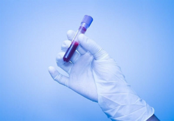
Because most neuro biomarkers are present in higher concentrations in cerebrospinal fluid (CSF), CSF analysis has been the best method of measuring relevant biomarkers. However, CSF sampling is a challenging and costly procedure that increases patient risk.
Reaching a sensitivity previously unattainable, Quanterix’s Simoa® NfL and GFAP immunoassays can reliably detect MS biomarkers directly from blood samples. Blood-based Simoa® immunoassays open the door to a quicker, non-invasive way to refine MS management, enabling early identification of high-risk patients who could benefit from more aggressive treatment options. In combination with traditional methods, Simoa® technology offers new perspectives for MS therapeutic management. Although only approved for research use, the capabilities of Simoa® have the potential to revolutionize how we approach, detect, and manage multiple sclerosis.
Conventional MS diagnostic methods: limitations for asymptomatic patients
The similarity in symptoms with other conditions make the diagnosis and staging of multiple sclerosis challenging. Clinicians need to perform several tests to ensure an accurate diagnosis and proper therapeutic management. MS diagnosis currently relies on examining a patient’s medical history, physical abilities, and potential cerebral lesions.
Magnetic resonance imaging (MRI) is the current gold standard for MS diagnosis.5 MRI radio waves reveal demyelinated lesions on the brain and spinal cord, allowing clinicians to monitor disease progression in the central nervous system. In addition, the Expanded Disability Status Scale (EDSS), developed to quantify MS-related disabilities, allows neurologists to determine the degree of severity for patients with clinically definite MS.3
Neuro biomarker detection in CSF already generates essential data for early MS diagnosis. However, newer technology aims to offer more accessible methods that move away from risky sampling procedures, including lumbar puncture.
Currently, there are no definitive blood tests for diagnosing and staging MS. However, progress in blood-based multiple sclerosis biomarker detection techniques offers promising insights for the future of MS management. Recent advances in NfL and GFAP detection are paving the way for accessible, quick, and reliable techniques to complement conventional imaging and scoring methods.4,6
Neurofilaments as biomarkers in multiple sclerosis: early demyelination detection
NfL is one of the most important biomarkers for predicting cognitive decline.4,6 Highly correlated with the degree of neuroaxonal damage, NfL level monitoring can predict the transition from the presymptomatic to the symptomatic stage, and offer valuable insights into disease progression.6,7
Because NfL is released into the CSF and bloodstream following axonal injury, it constitutes a powerful tool for characterizing in-progress neurodegeneration, including in MS.
Did you know? NfL concentrations in blood are reportedly 40-fold lower than in CSF, requiring highly sensitive technologies to ensure detection and quantification accuracy.
Quanterix’s Simoa® Nf-light™ assay kit is designed to detect NfL at ultralow levels, far below the Lower Limit of Quantification (LLoQ) found in typical immunoassays. Through this ultrasensitivity, Simoa® technology reliably detects NfL in serum and plasma and offers easy access to critical information.
Recent research using Simoa® technology reported higher serum NfL levels in MS patients compared with control individuals.8 Simoa® is an ultrasensitive and easy-to-use detection method that can simplify the identification of appropriate candidates for clinical trials and track treatment efficacy among MS patients.
Publication Highlight: Serum Neurofilament light: A biomarker of neuronal damage in multiple sclerosis
GFAP and astroglial activation: promising biomarker of MS progression and severity
GFAP is a strong candidate biomarker for indicating disease progression and severity in MS.9,10 Higher levels of GFAP have been found in MS patients with greater neurological disability, and these levels also correlate with different subtypes of MS.9,10,11 Although GFAP is less established than NfL in MS detection and staging, this astrocyte intermediate filament protein constitutes a hallmark of chronic MS lesions and represents a promising biomarker for indicating MS severity and monitoring disease progression.
Translating GFAP as a multiple sclerosis biomarker from research into clinical practice requires comprehensive characterization and testing. Ultrasensitive, large-scale biomarker detection methods could significantly accelerate GFAP’s validation and further implementation in clinical settings.
The Simoa® GFAP assay by Quanterix is an ultrasensitive high-throughput immunoassay that enables GFAP detection in CSF and blood samples with a level of sensitivity previously unattainable. By detecting GFAP directly in serum, Simoa® technology allows rapid data generation and repeat analysis in an easy, non-invasive way.
Did you know? Cohort studies using Simoa® GFAP as a readout reported that GFAP serum levels reflect MS disease severity better than CSF levels.12 Specifically, GFAP serum levels are associated with MS clinical severity scores and MRI lesion count, while GFAP CSF levels mainly correlate with disease duration.12,13 These results highlight the importance of novel detection methods utilizing MS biomarkers in comprehensive diagnosis and therapeutic management.
Publication Highlight: Glial Activation Markers In CSF And Serum From Patients With Primary Progressive Multiple Sclerosis: Potential Of Serum GFAP As Disease Severity Marker?

Exploring new biomarkers of multiple sclerosis: Future of MS research
Despite the promise of NfL and GFAP as MS biomarkers, a growing number of candidates correlating for axonal and neuronal damage await validation. For example, levels of the amyloid-precursor protein, known as APP, and tubulin beta (TUBβ) are reportedly higher in MS patients than in individuals with other neurological diseases.4
By evaluating differences in biomarker concentrations between cognitive conditions, biofluid-based detection methods can help current diagnostic tools distinguish MS symptoms from other neurodegenerative diseases. In addition, fluctuations in biomarker levels correlating with disease activity can provide important indications for therapeutic response.14
Through easy-to-use, accessible, and large-scale testing, blood-based biomarker detection methods can significantly boost biomarker validation and take MS detection to the next level.
Quanterix supports clinicians and researchers in improving MS detection by providing ultrasensitive tools to facilitate neuropathology research translation from discovery to the clinic.
Simoa® ultrasensitive immunoassays help:
- Detect MS biomarkers directly from blood samples, even at ultra-low levels
- Identify high-risk patients during the asymptomatic phase of MS
- Simplify candidate recruitment for clinical trials through an early patient screening
- Perform multiple or repeated MS biomarker analyses in a quick, cost-effective manner
- Accelerate validation of new MS biomarkers and help distinguish MS from other neurological diseases
- Predict therapeutic response and monitor effectiveness
Summary
MS disease progression and management represent a challenge to patients, researchers, and clinicians. Slowing MS progression and implementing appropriate treatment relies on the early identification of high-risk patients. With the ability to detect MS biomarkers through a simple blood test, Simoa® NfL and GFAP immunoassays allow quick, non-invasive, and cost-effective assessments of neuroaxonal damage. In addition, the ultrasensitivity of Simoa® enables early multiple sclerosis biomarker detection to facilitate clinical trial design and accelerate neuropathology research translation from discovery to the clinic.
Learn More About How Quanterix Simplifies Research in Neurology
References
1. Number of people with MS | Atlas of MS. Accessed February 13, 2023. https://www.atlasofms.org/map/united-kingdom/epidemiology/number-of-people-with-ms
2. Dobson R, Giovannoni G. Multiple sclerosis – a review. Eur J Neurol. 2019;26(1):27-40. doi:10.1111/ene.13819
3. Ziemssen T, Akgün K, Brück W. Molecular biomarkers in multiple sclerosis. J Neuroinflammation. 2019;16(1):272. doi:10.1186/s12974-019-1674-2
4. Yang J, Hamade M, Wu Q, et al. Current and Future Biomarkers in Multiple Sclerosis. Int J Mol Sci. 2022;23(11):5877. doi:10.3390/ijms23115877
5. Hemond CC, Bakshi R. Magnetic Resonance Imaging in Multiple Sclerosis. Cold Spring Harb Perspect Med. 2018;8(5):a028969. doi:10.1101/cshperspect.a028969
6. Gaetani L, Blennow K, Calabresi P, Di Filippo M, Parnetti L, Zetterberg H. Neurofilament light chain as a biomarker in neurological disorders. J Neurol Neurosurg Psychiatry. 2019;90(8):870-881. doi:10.1136/jnnp-2018-320106
7. Loeffler T, Schilcher I, Flunkert S, Hutter-Paier B. Neurofilament-Light Chain as Biomarker of Neurodegenerative and Rare Diseases With High Translational Value. Front Neurosci. 2020;14. Accessed February 14, 2023. https://www.frontiersin.org/articles/10.3389/fnins.2020.00579
8. Serum Neurofilament light: A biomarker of neuronal damage in multiple sclerosis – PMC. Accessed February 14, 2023. https://www.ncbi.nlm.nih.gov/pmc/articles/PMC5519945/
9. Axelsson M, Malmeström C, Nilsson S, Haghighi S, Rosengren L, Lycke J. Glial fibrillary acidic protein: a potential biomarker for progression in multiple sclerosis. J Neurol. 2011;258(5):882-888. doi:10.1007/s00415-010-5863-2
10. Meier S, Willemse EA, Schaedelin S, et al. Serum Glial Fibrillary Acidic Protein Compared With Neurofilament Light Chain as a Biomarker for Disease Progression in Multiple Sclerosis. JAMA Neurol. 2023;80(3):287–297. doi:10.1001/jamaneurol.2022.5250
11. Sun M, Liu N, Xie Q, et al. A candidate biomarker of glial fibrillary acidic protein in CSF and blood in differentiating multiple sclerosis and its subtypes: A systematic review and meta-analysis. Mult Scler Relat Disord. 2021;51. doi:10.1016/j.msard.2021.102870
12. Abdelhak A, Hottenrott T, Morenas-Rodríguez E, et al. Glial Activation Markers in CSF and Serum From Patients With Primary Progressive Multiple Sclerosis: Potential of Serum GFAP as Disease Severity Marker? Front Neurol. 2019;10:280. doi:10.3389/fneur.2019.00280
13. Abdelhak A, Huss A, Kassubek J, Tumani H, Otto M. Serum GFAP as a biomarker for disease severity in multiple sclerosis. Sci Rep. 2018;8(1):14798. doi:10.1038/s41598-018-33158-8
14. Akgün K, Kretschmann N, Haase R, et al. Profiling individual clinical responses by high-frequency serum neurofilament assessment in MS. Neurol Neuroimmunol Neuroinflammation. 2019;6(3):e555. doi:10.1212/NXI.0000000000000555